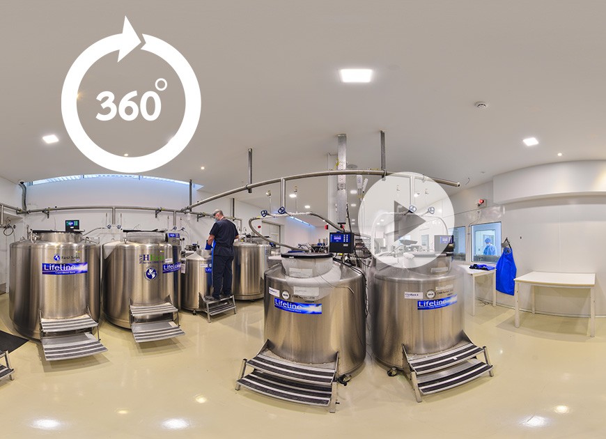
Virtual Tour Of LifeLine (VR-360)

We show that WJ has in vitro osteogenic differentiation capacity and in vivo, enhances bone growth in animal cleft palate models indicating its potential use as a natural tissue engineering construct for regenerative clinical applications. The success of this approach would represent a paradigm shift in the treatment of CLP patients by reducing or eliminating the need for subsequent bone grafting.
Greives, Matthew R. MD; Sahai, Suchit PhD; Wilkerson, Marysuna BA; Xue, Hasen BA; Teichgraeber, John F. MD; Cox, Charles MD; Triolo, Fabio PhD
Plastic and Reconstructive Surgery – Global Open: April 2017 - Volume 5 - Issue 4S - p 92–93
doi: 10.1097/01.GOX.0000516645.15717.06
PSRC 2017
PURPOSE: Secondary alveolar bone grafts are the standard treatment for patients with cleft lip and palate (CLP), but remain invasive and have several disadvantages such as delayed timing of alveolar repair, donor-site complications, graft resorption and need for multiple surgeries. Earlier management of the alveolar defect would be ideal, but is limited by the minimal bony donor sites available in the infant. Wharton’s Jelly (WJ), the connective tissue matrix of the umbilical cord, is a gelatinous substance comprised of proteoglycans, collagen, and is rich in perinatal stem cells. Our hypothesis is that inclusion of WJ in the alveolar pocket of CLP patients at the time of palate repair enhances bone growth and accelerates healing, with the autologous stems cells and extracellular matrix serving as a “primary bone graft” to close the alveolar cleft.
METHODS: Human umbilical cords were harvested following routine delivery and WJ was isolated and purified. In vitro, WJ derived stem cells were placed in osteogenic differentiation medium for 14 days, followed by Alizarin Red S staining to evaluate mineral deposition. In vivo, we used a rat critical-size alveolar bone defect model to investigate the use of Wharton’s Jelly (WJ) in formation of bone. WJ was implanted into a critical size (7 x 4 x 3 mm) alveolar bone defect model representative of cleft palate surgery in 10–11 week old male Sprague-dawley rats. The defects were monitored weekly with CT imaging of living animals to evaluate bone formation in time, followed by histology evaluation at week 24.
RESULTS: WJ showed significant in situ osteogenic differentiation of WJ cells as evidenced by strong Alizarin Red S staining. By contrast, no staining is observed when nWJ is maintained in control medium. In vivo, CT data showed that the defect size was critical and did not lead to the union of the bones in the control animals (n=12) for the entire duration of study. New bone growth was stimulated leading to partial-to-full closure of the defect in the animals treated with WJ (n=12). 24 weeks postoperatively, the percent increase in new bone formation in the WJ treated group (156.57 ± 26.85%) was markedly higher than that in the control group (49.97 ± 12.51%) (p<0.05). Histology data also revealed significantly greater new bone formation in WJ treated vs, control animals, confirming CT findings.
CONCLUSION: We show that WJ has in vitro osteogenic differentiation capacity and in vivo, enhances bone growth in animal cleft palate models indicating its potential use as a natural tissue engineering construct for regenerative clinical applications. The success of this approach would represent a paradigm shift in the treatment of CLP patients by reducing or eliminating the need for subsequent bone grafting.
LFLN REF:02112017,p.8