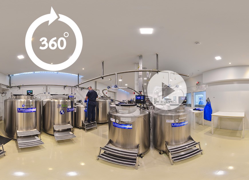
Virtual Tour Of LifeLine (VR-360)
Putnam Valley, NY. (Mar. 6, 2014) – When human umbilical cord blood cells were transplanted into rats that had undergone a simulated myocardial infarction (MI), researchers investigating the long term effects of the transplantation found that left ventricular (LV) heart function in the treated rats was improved over those that did not get the stem cells. The animals were maintained without immunosuppressive therapy.
The study will be published in a future issue of Cell Transplantation but is currently freely available on-line as an unedited early e-pub at: http://www.ingentaconnect.com/content/cog/ct/pre-prints/content-ct0860Chen.
"Myocardial infarction induced by coronary artery disease is one of the major causes of heart attack," said study co-author Dr. Jianyi Zhang of the University of Minnesota Health Science Center. "Because of the loss of viable myocardium after an MI, the heart works under elevated wall stress, which results in progressive myocardial hypertrophy and left ventricular dilation that leads to heart failure. We investigated the long term effects of stem cell therapy using human non-hematopoietic umbilical cord blood stem cells (nh-UCBCs). These cells have previously exhibited neuro-restorative effects in a rodent model of ischemic brain injury in terms of improved LV function and myocardial fiber structure, the three-dimensional architecture of which make the heart an efficient pump."
According to the authors, stem cell therapy for myocardial repair has been investigated extensively for the last decade, with researchers using a variety of different animal models, delivery modes, cells types and doses, all with varying levels of LV functional response. They also note that the underlying mechanisms for improvement are "poorly understood," and that the overall regeneration of muscle cells is "low."
To investigate the heart's remodeling processes and to characterize alterations in the cardiac fiber architecture, the research team used diffusion tensor MRI (DTMRI), used previously to study myofiber structure in both humans and animals.
While most previous studies have been focused on the short term effects of UCBCs, their study on long term effects not only demonstrated evidence of significantly improved heart function in the treated rats, but also showed evidence of delay and prevention in terms of myocardial fiber structural remodeling, alterations that could have resulted in heart failure.
When compared to the age-matched but untreated rat hearts with MI, the regional myocardial function of nh-UCBC-treated hearts was significantly improved and the preserved myocardial fiber structure may have served as an "underlying mechanism for the observed function improvements."
"Our data demonstrate that nh-UCBC treatment preserves myocardial fiber structure that supports the improved LV regional and chamber function," concluded the researchers.
"This study provides evidence that UCBCs could be a potential therapy with long term benefits for MI" said Dr. Amit N. Patel, director of cardiovascular regenerative medicine at the University of Utah and section editor for Cell Transplantation. "Preservation of the myocardial fiber structure is an important step towards finding an effective therapy for MIs"
Source: Cell Transplantation Center of Excellence for Aging and Brain Repair
LFLN REF. 27.03.14 P.34

The facility of CBB Lifeline Biotech is Accredited by AABB International Standards for Cellular Therapy Products for Cord Blood Banking Services

Since October 2020 the clinical testing is performed by a third party accredited collaborator


LifeCord Patented Methodology
US Patent Office